Project timelines and workflow are adversely impacted by the amount of assay development required for each protein when using traditional flow-cell based or ELISA based technologies. Several factors contribute to delays in assay development, such as limited throughput, sample constraints due to fluidics or serial dilution, parallel processing, and particularly for ELISAs, identification of the appropriate capture and detection antibody.
In direct contrast, the Octet® family of instruments facilitates method development in kinetics and custom protein quantitation assays. Eight-channel parallel processing allows for multiple conditions or molecule types to be assayed for direct side-by-side comparison, streamlining the assay development process.
pH Scouting
The ideal immobilization pH is slightly below the protein's isoelectric point (pI). If the pI is unknown, then a pH scouting experiment can quickly determine the optimal pH that effectively immobilizes the ligand to the amine reactive biosensor surface. The data is examined for the maximum signal relative to a negative control.
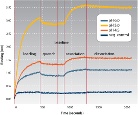
Figure 1: Real-time binding chart of pH scouting of immobilized HA-GST to an interacting antibody.
Using the Amine Coupling Reagent Kit, HA-Tagged GST (10 µg/mL) was prepared in 100 mM MES Buffer at pH 4.5, 5 and 6 and immobilized onto Amine Reactive Biosensors using the Octet QK System (Figure 1). The association and dissociation of an interacting antibody (20 nM) to the immobilized ligand was assayed. The sample at pH 5 exhibited maximum signal for both loading and association steps relative to the other conditions and was selected for future kinetic analyses.
Regeneration Buffer Selection
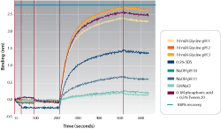
Figure 2a: Real-time binding chart of regeneration conditions run in parallel on the Octet QK System.
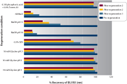
Figure 2b: Percent recovery evaluation of regeneration conditions run in parallel on the Octet System.
Regeneration on the Octet System allows for reuse of the biosensor tips for subsequent analysis with the immobilized protein. Figure 2a exhibits the real-time binding chart of eight regeneration buffers evaluated in parallel. Each set of eight biosensors was regenerated and reused for another interaction. Figure 2b illustrates the percent recovery of a protein to a regenerated ligand surface. The regeneration conditions that show close to 100 percent recovery after repeated regenerations are good candidates for further development. The ability to test regeneration reagents in parallel facilitates rapid assay development time for the protein of interest.


